-
2020
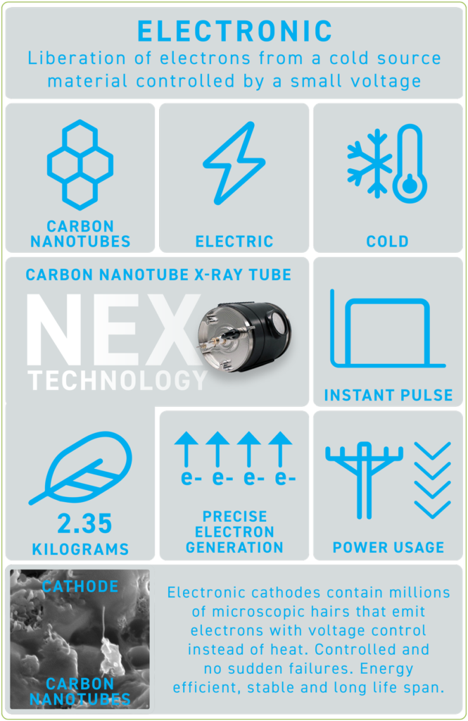
ELECTRONIC
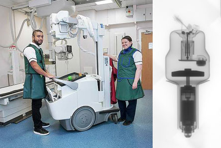
DIGITAL RADIOGRAPHY
NEX TECHNOLOGY uses less power, emits minimum heat with no need for oil cooling. Lighter, simpler & more reliable system with no glass, filaments or rotating anode. -
1980s
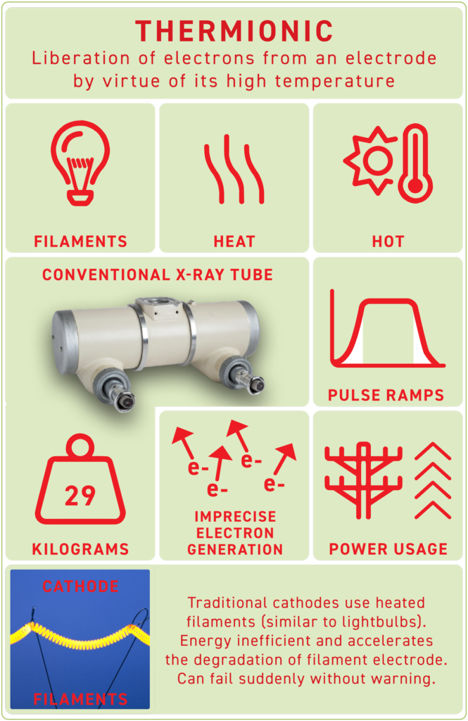
THERMIONIC
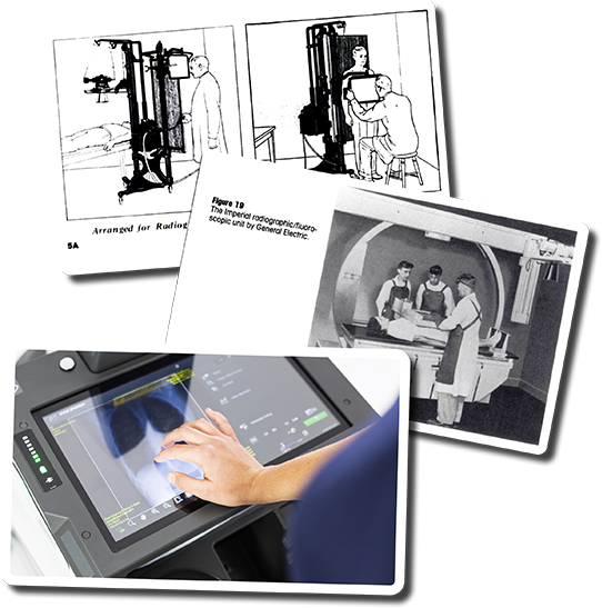
COMPUTED RADIOGRAPHY
The use of computers required no chemicals, had faster processing time, created digital images stored on hard drives. Can be corrected for over/under exposure & rotation. -
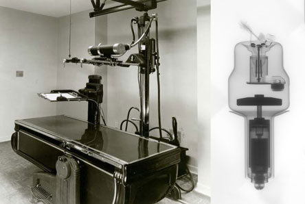
1933
THERMIONIC
ANALOGUE
Rotating anode tubes helped improve the speed and efficacy of X-rays, and their use was broadened. -
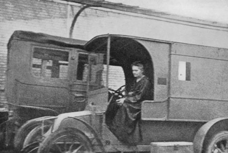
1917
THERMIONIC
ANALOGUE
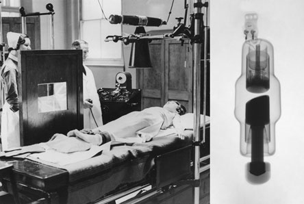
WW1 – Marie Curie invents first “radiological car” with X-ray machine & darkroom for surgeons. -
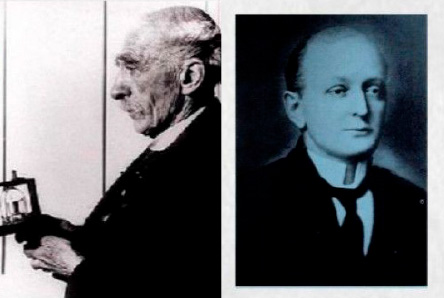
1904
THERMIONIC
ANALOGUE
John Ambrose Fleming invents the vacuum tube. Clarence Dally dies due to exposure to radiation. -
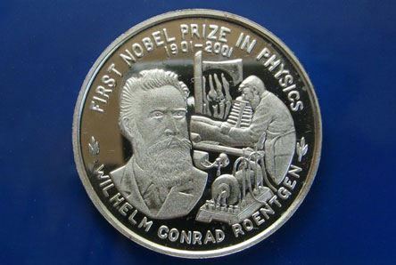
1901
Wilhelm Conrad Roentgen, won the first Nobel Prize for Physics for discovering the x-ray in 1895.
-
2000s
THERMIONIC
DIGITAL RADIOGRAPHY
Instantaneous digital images display with higher resolutions. Lower doses of x-ray exposure. -
1940s & 50s
THERMIONIC
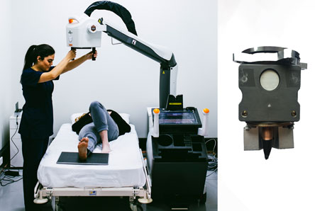
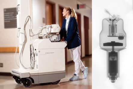
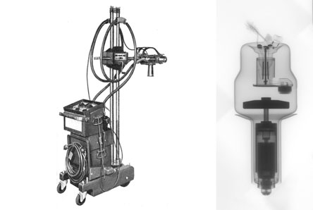
ANALOGUE
X-rays became increasingly useful in treatment and diagnosis of serious issues. More safety regulations and precautions were put in place. Mobile X-rays developed. -
1922
THERMIONIC
ANALOGUE
200,000-volt x-ray tube allowed radiographs of thick steel parts to be produced in a reasonable time. -
1903
THERMIONIC
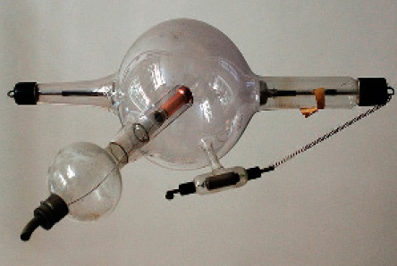
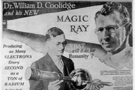
ANALOGUE
William Coolidge invents the x-ray tube, which made possible the continuous emission of x-rays. -
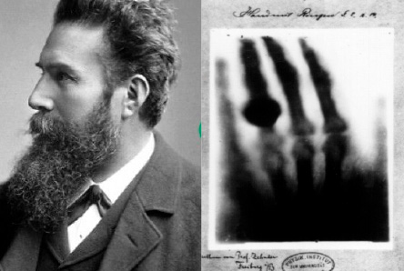
1895
THERMINONIC
ANALOGUE
Wilhelm Roentgen’s first research into mysterious ‘x-rays’. Soon after he conducts the first x-ray on his wife’s hand.